Please remember!

The information presented on this blog represents "learning in progress" on my part, a horse owner, who was not satisfied with professional farriers and took matters in my own hands. As far as I am aware at the time of the post, the information presented is correct, but may change with me understanding more about hooves, in which case I will edit or remove the post. In order to follow my learning and understand everything about Molly's hoof, you need to start reading at the bottom.

Thursday, October 18, 2012

"geometric" vs. "result-oriented"


This paper describes the two methods that I used to balance Molly's front right hoof:

***********************************************************************************
http://www.ncbi.nlm.nih.gov/pubmed/1874681

J Am Vet Med Assoc. 1991 Jun 1;198(11):1980-9.

Factors involved in the balancing of equine hooves. 

Balch O, White K, Butler D.

Department of Veterinary and Comparative Anatomy, Pharmacology and Physiology, College of Veterinary Medicine, Washington State University, Pullman 99164.

***********************************************************************************

The two methods are: the "t-square (geometric)" method and the "result-oriented" method.

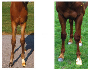
The 2 pictures below are taken from that paper. The case shown is very similar to Molly's case, "base-narrow and toed-out". And this horse too lands on his lateral quarter/toe just like Molly does.

This sentence was especially encouraging:

"Use of Moyer and Anderson's result oriented technique is often more successful in treating lameness than the geometric procedure."

If it treats lameness better, it must be better for Molly, who thankfully never was lame.


Wednesday, October 17, 2012

balancing Molly's front right hoof


This topic is the reason for this whole effort with the blog.


I wanted to give an as comprehensive overview of what I have observed so that I can get additional suggestions with respect to Molly's FR balance.

As I have shown, the leg is crooked. This crookedness has been my major struggle with knowing how to balance exactly.


hindleg conformation

I have no pictures of my horses hindlegs but I have looked at them often and found that just like the forelegs are slightly valgus, so are the hind legs. In the case of hind legs one often speaks of cow-hocked.  Here again, the "textbook" knowledge indicates that straight legs are ideal. However, I found this picture on the web:


This is an excerpt from a book:


It states: "Most breeders would add cow hocks to the list, but this is incorrect. Horses are differentiated from other mammals precisely on the basis of the fact that horse hock bones (and stifle joint structure) force them to stand with their hocks pointing inward. In other words, a horse that is not cow-hocked is not horselike.; in fact, a horse with "straight" hocks as seen from the rear (figure 32b) is likely to move with wobbly, pathology-generating hocks. There are degrees of cow hock: if extreme enough to cause interference (figure 32c), the fault is a serious one, but hocks that face in are not a fault per se."

I am reasonably convinced, my horses hind legs conformation is absolutely fine, normal and healthy. They look just like picture 32A above, maybe even a bit straighter than this example.


landing patterns

I made this slide to show how Molly is walking. As a consequence of the leg conformation, she has the tendency to impact the ground with her lateral side first. It is particularly obvious on her FR, the one foot that has the offset P1 (long pastern bone), where Molly most of the time hits the ground with her lateral toe first. The FL lands flatter, but also lateral side first.


forleg conformation

I have tried to compare the front and hind leg conformation of my horses to the "textbook" examples.

The conformation that is considered ideal is straight legs, defined as:

"Ideally, when viewing the forelegs from the front, a straight line from the point of the shoulder should bisect the entire column of bone all the way to the toe, with equal portions of the bone on either side of the bisecting line."

 From: http://www.extension.org/pages/10255/relating-form-to-function:-horses-frontlegs-front-view


Both my horses definitely do NOT have straight legs like those above! Their front legs seem to be slightly knock-kneed (also called valgus).

Gus front leg conformation

Molly front leg conformation

Several pieces of evidence suggest that a slight valgus leg conformation is actually normal, if not beneficial.

1) This website states that a slight valgus conformation is normal.
2) This study based on injuries of racing Thoroughbreds states: "An increase in the carpal angle as viewed from the front (carpal valgus) may serve as a protective mechanism, as the odds for a carpal fracture and carpal effusion decreased with an increase in the carpal angle."


When I pick Gus' legs up and bring them forward and look down the cannon bone from the knee, the legs down from the knees are completely straight.  The same is true for Molly's left front. Molly's right front, her problem foot with the side bone, however is different: the cannon bone, and the short pastern are "offset". This can be best seen on the x ray.


 See this picture for terminology of bones:


Molly's offset long pastern bone is most likely something that has developed as a foal, due to bad hoofcare and/or injury of some sort. Today, at 8 years of age, this is nothing I can change. Molly's hoof has obviously suffered from this leg conformation, as she developed side bones in this hoof but not in her other front hoof. I am reasonably sure that she had this side bone already as a 3.5 year old, as I remember specifically asking the vet about that bulge that she had in her coronary when I bought Molly. The vet at that time said it was nothing to be concerned about. I guess he is correct, Molly has arranged herself with the situation, she has never been lame, but her FR is a long term liability and I want to do everything in my power to at least provide it with the best balanced trim given her leg conformation. So far, everything looks quite good, the joint spaces are relatively regular and to the degree that this rather bad x-ray can tell, there is no sign of calcifications (arthritis). Not yet! My goal is to keep it this way.

Monday, October 15, 2012

Natural asymmetry and hoof wear

The most important skill of a trimmer is to observe the horse during movement and interpret the pattern of hoof wear.

Early on, before I knew anything about the natural asymmetry, I observed that both my horses showed more wear on their lateral sides. The white line always seemed more separated on the lateral than the medial side.

This is pretty easy to explain now, as the lateral side receives more pressure during movement it also receives more wear, especially at the toe, as the hoof rolls over that lateral toe more than the medial when leaving the ground.


In the beginning I misinterpreted the fact that the white line separation was wider and deeper laterally and lowered the lateral wall. This was a mistake, that I luckily caught in time, before the hoof looked like these:


The hooves in the picture above also shoes a wider lateral side than medial, but this has nothing to do with "natural asymmetry" as now the lateral walls drift away and the coronary band is no longer straight but curves down medial-laterally. This asymmetries has to be distinguished from the natural asymmetry. 


In summary, the principles of natural asymmetry of the hoof and the patterns of locomotion as detailed in the previous entries of this blog requires hoof care practitioners to balance the surplus of wear on the lateral side by adjusting the medial side if necessary.


Natural asymmetry and locomotion

The hoof has to fulfill two important functions. It needs to carry the weight of the horse, and it needs to balance the horse during locomotion, when at some points in time, the horses' weight is carried by a single hoof.

The illustrations below are referenced from this very informative website.

The static load is distributes equally on both front legs:
The dynamic load however can be concentrated on either one of the two front legs:

In order to balance the weight of the front half of the horse on a single hoof, the horse loads the lateral side 10-20% more:


I have found, that the horse performs this "balancing act" by moving the leg slightly towards the mid-line.

My horse Gus provides a good example for this. He has rather straight legs (very slightly base narrow) and I have not seen any horse with more healthy and regular hooves than his, yet he places his front feet in such a way that they load the lateral side more, by placing them towards the midline. This specific way of walking is more pronounced in soft ground, most likely as there is more need for balancing the weight on soft ground. But it also can be seen on harder ground.  On a level surface, like a tarmac road, Gus lands heel first or flat and this placement of the legs towards the midline is not very obvious, but to some degree it can be seen even on a hard flat surface.

It is interesting to note that the same conclusion has been drawn by the author of this website:
http://otherideas.typepad.com/christine_sander_hufbalan/

The article is in German, but the first picture shows this specific way of advancing the legs towards the midline.


Taken together these observations suggest (as has been done here) that the lateral side of the horse's hoof is wider and flatter because it is the "balancing side" of the hoof, and the medial side is primarily involved for the static load bearing of the leg.

It may also be worth noting that as the weight of the horse increases, so is the need to balance the horse's weight during locomotion, when temporarily only one leg carries the weight. The heavier the horse, the more balancing on the lateral hoof side is required.

Natural asymmtery - fossil horse bones

The ancestor of all mammals (and thus the horse) had five digits on each limb involved in locomotion. The horse, while still having remnants of all 5 digits, has reduced 4 of them and only uses one for locomotion: the middle digit (III), equivalent to the human middle finger. The reduction of the weight bearing digits was gradual during horse evolution, as can be seen here:


As an evolutionary biologist, I find it interesting to look at the evolution of the bones, changes that occurred represent mainly adaptations of the horse lineage to its environment, and also developmental constraints.

I was very excited to see this study. The authors report a well-preserved skeleton of a 4.6 million-y-old three-toed horse (Hipparion zandaense) from the Zanda Basin, southwestern Tibet.

Hipparion zandanese must have looked like this:


 In the supplemental material of the PNAS paper, they show actual pictures of fossilized (4.6 Million years old) bones.


Based on what I know about the horse's hoof, I think the Figure legend must be a mistake, this is not the right front foot, but the left, based on the shape of the coffin bone. The coffin bone of this 4.6 Million year old ancestor of the modern horse shows a clear asymmetry,  that is also characteristic of the modern horse, including MINE! Most likely the steeper and slightly smaller medial side is adaptive for the horse, exactly why I have not yet figured out, but will try and find answers. The answer will lay in the biomechanics of the hoof.

trimming toe sole

This entry on the Easycare blog is important for me. As my horses develop that excess sole in the toe, especially on their hinds. I have been doing exactly as this person describes, rasp flat over that excess sole, until the collateral grooves at the tip of the frog come down to about 9 mm.


Wednesday, October 10, 2012

Sidebone

Molly has sidebone on her FR, it is pretty obvious to see on her X ray and also from directly looking at her hoof. Today, someone posted in a hoof forum a picture of a dissected foot showing side bone. Molly's are not as advanced but still, it is quite amazing to actually see them being elevated beyond the capsule. I can definitely feel them on Molly's FR. Also, her hoof has a similar triangular shape, just like this one.


I have ordered this balm and will apply it to her coronary band, according to this short movie:


I am not particularly hopeful that this is going to make a huge difference but on the other hand, if bone can demineralize as in osteoporosis, why should cartilage not demineralize too, once the hoof is properly trimmed and correct hoof mechanics restored. I see Molly itching herself often just underneath those ossifications.


Molly's sidebone can be seen here. It is more pronounced on the lateral side. But she does have it on both sides of her hoof. Her front left does not have any signs of side bone. Her front left is such a healthy hoof, it is a real joy to look at it!


Sunday, October 7, 2012

Biomechanics of the hoof - natural asymmetry

I have spent literally hours trying to understand my horses' leg conformation, and how it affects their movements and ultimately hooves. This is a whole area of hoof care I have not yet known much about. The discipline itself is called "Biomechanics of the Hoof".

Unfortunately, every horse is an individual and thus general rules are hard to formulate. But there are SOME rules that are worthwhile knowing. Below I summarize my very limited understanding of this topic, I will keep updating it as I learn and understand more.

1.  Natural asymmetry

Most healthy hooves show a pattern of "natural asymmetry", which describes the fact that the medial (inside) hoof wall is usually steeper than the lateral (outside) hoof wall. Here is an theoretical example, taken from an article by James Rooney, D. V. M.:


 As a consequence, the side of the hoof with the steeper wall is usually slightly smaller. This can be seen from this picture of a dead mustang on the road (origin of picture unknown). The yellow and the orange line have the same length.


Brian Hampson, in his study of several populations of Australian wild horses (Brumbies) inhabiting various different habitats, found that all populations apart from one (the desert rocky population, which is not a natural and ancestral habitat for the horse!) showed the same pattern of a steeper medial than lateral hoof wall (see page 44 of Peter Ramey: Care and Rehabilitation of the Equine foot). So this observation seems to be a real and general one, and as such demands an explanation.

According to James Rooney, this pattern is a direct consequence of the forces that act on the hooves by the "center of mass" of the horse, which is concentrated in the middle, i.e. between the two hooves. The ratio of the "outward" to "downward" vector of the forces exerted by the body mass of the horse is skewed towards outside on the lateral hoof walls, thus shaping the hoof in this way.


This model would predict that the medial wall would be falling behind the vertical, something that is not usually observed. I think this tendency is offset by the way the legs advance on a healthy, straight-legged horse. I will come back to this aspect in a new post, describing how a healthy horse loads its hoof during movement.

Newborn foals do not show such a medial-lateral wall angle asymmetry. It only develops once the hoof is subjected to the body weight of the horse.

My horses both show the phenomenon, though Gus much less pronounced than Molly. One explanation for that fact could be that Gus grew up on hard rocky ground (in Texas), similar to the desert rocky brumbies,  and Molly on soft (wet) pastures in Michigan. The dry, rocky conditions in Texas have formed a hoof that has more similar steep walls medially and laterally and as the coffin bone grows it is shaped by the hoof capsule around it to take on that same pattern.


We have to assume that in order for the previous principles to apply the horse must have healthy normal straight (slightly valgus can be considered normal, see picture below) legs.

This slight valgus leg conformation can be considered normal
There are numerous conformational abnormalities, that would change the above explanations as rotational forces come into play. This is precisely the reason what makes the study of the biomechanics of the hoof so difficult, at least for me!